Home / Treatments / Ankle: arthroscopy & ligaments
The sprain that "never healed properly" is real — even when the scans look fine. It has a name, an anatomical explanation and a solution.
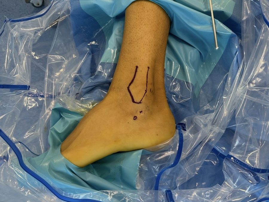
The ankle sprain is sport's most common injury — and its most underestimated one. Most heal well with proper functional treatment, but a significant number of people develop chronic instability: repeated twists on minimal provocation, a feeling of giving way, persistent pain, or no confidence on uneven ground. And there is a second, subtler picture that leaves many people with pain and "normal" scans for months: microinstability. To understand both, we need to look closely at the ligaments.
For decades the anterior talofibular ligament (the one injured in most sprains) was described as a single structure. The anatomical studies by Vega and Dalmau-Pastor's group showed something different — and clinically decisive: this ligament has two fascicles, present in 100% of the ankles studied, and they don't behave the same way:
The superior fascicle (red) lives inside the joint; the inferior fascicle and calcaneofibular ligament (blues) form one connected complex.
Isolated tear of the intra-articular superior fascicle.
Injury of the full complex (inferior fascicle + calcaneofibular ligament).
Both can be treated — but the first, precisely because it hides from the scans, is the one that leaves so many people with a "badly healed sprain" for months or years. Suspect microinstability when you see:
The first step is always conservative: physiotherapy with strength and proprioception work. When symptoms persist, arthroscopy does two things at once: it confirms the injury by seeing it directly — often invisible on imaging — and treats it in the same procedure.
Ankle arthroscopy works through millimetre incisions with a high-definition camera, treating the inside of the joint with minimal tissue damage. When instability is the problem, anatomic ligament repair restores tension to your own ligaments with small anchors; both techniques are frequently combined in the same operation. Surgery takes place at IMED Elche or IMED Alicante hospitals, usually as a day case or short stay.
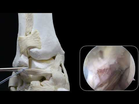
How the lateral ankle ligament is repaired with anchors (Arthrex technique). Plays here on click, in YouTube's no-cookie mode. Open on YouTube.
The anatomical work by Vega and colleagues (32 ankles dissected plane by plane) laid the foundations of modern ankle ligament surgery:
Its practical consequence: because the whole complex is connected, isolated arthroscopic repair of the anterior talofibular ligament gives excellent results even when the calcaneofibular ligament is also injured — and the arthroscopic technique is biomechanically equivalent to open surgery, with less tissue damage.
Protection in a splint or boot, swelling control and early protected motion depending on the procedure.
Progressive weight-bearing in the boot; physiotherapy begins: motion, strength and basic proprioception.
Normal gait, advanced strength and specific proprioception; progressive running once criteria are met.
Change of direction, sport-specific drills and return to sport by objective stability and strength criteria.
Acute sprains almost never do. Surgery is considered after repeated sprains with correct rehab when instability persists, or when there are associated injuries (osteochondral fragments, impingement).
The superior fascicle is a thin intra-articular structure that often goes unnoticed on conventional imaging. Its tear causes pain and low confidence without obvious instability — the typical picture of microinstability.
Microinstability is the isolated tear of the superior fascicle (subtle symptoms); classic instability is injury of the full complex (frank giving-way). Both can be treated.
Anatomic repair of the lateral ligaments (Broström-type) with small anchors — minimally invasive open or arthroscopically assisted, treating intra-articular injuries at the same time. If the calcaneofibular ligament is also torn, two repairs are not always needed: repairing the anterior talofibular ligament tensions the whole connected complex. Selected cases need augmentation or graft.
Damage to the cartilage and bone of the talar dome, often after sprains, causing deep pain and sometimes catching. Treated conservatively or arthroscopically depending on size and location.
As a guide: a few weeks of protection, progressive loading, and return to sport between 3 and 6 months depending on the procedure and the objective criteria achieved.
A specific examination and an in-clinic ultrasound scan point to the problem at the first visit — even if your scans "look fine".
Or by phone: +34 681 87 65 04
