Home / Treatments / Bunion surgery
When a bunion hurts and limits you, the bone is corrected. Today, through incisions of a few millimetres, walking the same day.
A bunion, or hallux valgus, is not a bone that grows: it is a progressive deviation. The first metatarsal drifts inwards and the big toe leans towards the other toes, so the head of the metatarsal sticks out on the inner side of the foot. That is the "bump" that rubs against the shoe.
There is a clear hereditary tendency (foot shape runs in families), and narrow or high-heeled shoes, flat feet and joint laxity speed the deformity up. Over time the big toe stops pushing off when you walk, the load shifts to the neighbouring metatarsals and calluses and pain under the sole (metatarsalgia) and hammer toes appear, most often in the second toe.
Left, a normal forefoot. Right, hallux valgus: the first metatarsal drifts inwards, the big toe leans outwards and the head of the bone sticks out: that is the bunion.
Bunions are not operated on for cosmetic reasons. Surgery aims to remove the pain, give the big toe its function back and stop the damage to the rest of the forefoot. It is considered when:
The decision is made in consultation with an examination of the foot and weight-bearing X-rays (taken standing), which show the true deformity and allow the correction to be planned.
Percutaneous or minimally invasive surgery (MIS) corrects the deformity through incisions of two to three millimetres, without opening the joint. Under X-ray guidance in the operating theatre, the first metatarsal is cut and shifted into its correct position (an osteotomy), the big toe's phalanx is corrected when needed, and the correction is held with percutaneous screws. It is performed under a regional anaesthetic block of the foot with sedation, and you go home the same day.
Before: the deviated bone and the cut line. After: the head is shifted outwards, under the toe, and held with a screw. All through 2 to 3 mm incisions.
Osteotomies through 2 to 3 mm incisions, fixed with screws.
An incision of several centimetres with direct correction of the deformity.
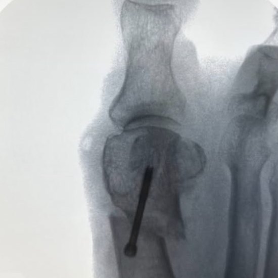
Intra-operative X-ray at the end of the correction: the head of the first metatarsal repositioned and held with a percutaneous screw.
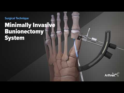
The keyhole bunion technique with screw fixation (Arthrex system). Plays here when you press it, in YouTube's cookie-free mode. Open on YouTube.
These timings are a guide: every foot and every person has their own pace, and they are adjusted at the check-ups.
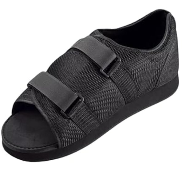
The post-operative shoe we use: a flat, rigid sole to bear weight from day one, an open toe and two velcro straps that adjust to the dressing. Worn for the first weeks.
You walk from the start in a stiff-soled shoe that protects the correction. Foot elevated as much as possible and painkillers on a schedule.
The bandage is part of the treatment: it holds the correction while the bone heals. Short distances only, and no normal shoes yet.
Control X-ray. Depending on healing, you move to a wide, comfortable shoe and start mobilising the toe.
Long walks, working on your feet and, later on, sport. Swelling of the foot can take several months to settle completely; that is normal and does not mean something is wrong.
The same technique, with tiny incisions and early weight-bearing, is used for other common foot problems, often in the same operation as the bunion:
No. They ease the pain and reduce rubbing, but they do not move the bone back into place. When the bunion hurts and limits you despite those measures, surgery is the only way to correct it.
It is done under a regional block of the foot with sedation, so the operation itself is painless. Afterwards, discomfort is managed with standard painkillers and is usually less than after open surgery because of the size of the incisions.
The same day, in the post-operative shoe. For the first weeks, short distances and the foot elevated; normal footwear returns progressively, guided by the check-up and the control X-ray.
In selected cases yes, because you bear weight from day one. It is decided in consultation; it is often more comfortable to operate on the more painful foot first and the other a few months later.
There is a small risk of recurrence, higher if not every component of the deformity is corrected or if narrow shoes are resumed. A complete correction planned on weight-bearing X-rays reduces that risk.
At the first visit the foot is examined, weight-bearing X-rays are taken and we decide whether surgery is worth it and with which technique.
Or by phone: +34 681 87 65 04
